|
|
|
|
|
Toll Free: (888) 768-6310 • International: +1 (615) 826-0582
|
|
|
|
|
|
|
PORTABLE ULTRASOUND >> Portable GE Ultrasound >> GE Systems
|
|
 |
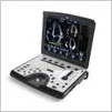
GE Vivid q
 A new advance in portable echocardiography, the Vivid q extends the reach of the medical professional to diagnose cardiovascular anatomy and LV function by adding more quantitative tools and pushing the boundaries to the highest levels of image quality. Vivid q builds on the many industry leading features and technologies of the Vivid i with more performance, new quantitative analysis tools and a wider range of applications.
The addition of the M4S-RS matrix array transducer enhances 2D images for adult cardiac applications.
Coded Octave imaging takes image quality to a new level by enhancing your images for Color Flow and Doppler Imaging.
Smart Depth automatically adapts imaging parameters to help newer users, as well as expert sonographers, see better results.
Smart Stress improves workflow, shortens optimization time, and provides reproducibility for review, wall-segment scoring, and reporting.
Parametric imaging and quantification support clinical decisions. The GE Vivid q builds on the many industry leading features and technologies of the Vivid i with more performance, new quantitative analysis tools and a wider range of applications.
Auto EF provides the most commonly used parameter to describe the LV function, the Ejection Fraction (EF). Auto EF provides the most commonly used parameter to describe the LV function, the Ejection Fraction (EF), which assists in finding the endocardial border. Auto EF also reduces the dependency of "seeing" the border in each image by analyzing and tracking the myocardial tissue. This feature also automatically locates the end systolic and end diastolicframes.
Automated Function Imaging (AFI) supports LV function analysis by visualizing the longitudinal wall, shortening, lengthening and highlighting a segment's contraction. AFI can potentially be used to differentiate disease from non-disease segments. It also decreases LV function assessment variability which provides clinical decision support, streamlining workflow.
Tissue Velocity Imaging (TVI) and Tissue Tracking (TT) show tissue velocity and displacement in the direction of the ultrasound beam with high temporal resolution.
Tissue Synchronization Imaging (TSI) translates comprehensive quantification into an easy-to understand image demonstrating mechanical synchronicity of different myocardial segments.
Intracardiac Echo (ICE) Imaging. By combining exceptional detail and quality of information with advanced interventional echocardiography ultrasound ICE imaging technology, the Vivid q opens up a "new window" to the heart. The Vivid q ICE catheters deliver excellent image quality and real- time visualization of cardiac structural anatomy with therapy catheters for monitoring and guidance during interventional procedures. ICE gives you a better understanding of structural orientation during trans-septal puncture procedures help avoid clinical complications.
OB/GYN Software
*NOTE* Not all options/features listed above are standard on the Vivid q®. |
|
|
|
|
|
 Ask for a Quote Today! Toll Free: 888.768.6310 Ask for a Quote Today! Toll Free: 888.768.6310
International: 615-826-0582 You will ALWAYS speak with an owner, not a commissioned sales person |
|
|
|
|
|
|
|
|